

Watch cancer spread cell by cell
A method that images cancer at the single-cell level has been developed by researchers in Japan. The technique uses chemical markers that make whole mouse bodies and organs highly transparent. When used in conjunction with current imaging methods they could get sufficient resolution to cancer cells multiplying within organs.
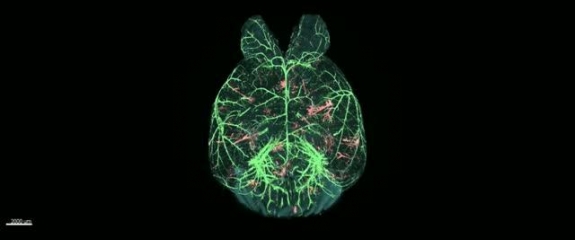
The imaging technology worked on lung, intestinal and hepatic tissues and could even identify nascent tumour cells as they traveled through the body to and from new tumours. The work, which appears in the July 5 issue of Cell Reports demonstrates the power of whole-body and whole-organ clearing and imaging with single-cell resolution. The technique can reveal cancerous colonies in sufficient detail to allow the calculation of their volume and distribution as well as the shape of the colony — all characteristics critical to distinguishing between patterns of metastasis. The researchers used the platform to generate a whole-mouse scan of pancreatic cells spreading into the liver and throughout the abdomen. Another set of images shows a healthy pair of lungs being colonized by cancer cells over the course of two weeks. Another probed the relationship between cancer cells and blood vessels inside the brain.
Capturing images of individual cancer cells during metastasis is challenging. They are often scattered throughout the body and locating them relies on detecting signals from fluorescent proteins that they express. These signals need to be preserved when applying tissue clearing methods in pursuit of higher resolution. The method employs a chemical cocktail termed CUBIC (Clear Unobstructed Brain/Body Imaging Cocktail) previously developed by the reporting team for whole-body imaging preparation. The team manipulated the composition of the cocktail to make the tissues and vital organs appear clear and allow them to better detect and view cancer cells. The researchers were able to pluck out fluorescence signals and locate cancer cells in places such as the liver, pancreas, and intestines.
The new imaging protocol is already providing a clearer view of certain mysterious steps of metastasis. Before settling at a new site in the body metastatic cancer cells travel through the bloodstream, entering and exiting through blood vessel walls. It seems that most cells do not survive the trip. Images developed with this new method suggest that cells treated with TGF-beta, a protein that regulates cellular growth and differentiation in humans and is produced in increased quantities by some cancers, are far more likely to survive the journey and form new malignant colonies.
The researchers feel that cancer imaging and analysis using their CUBIC protocol will lead to further insight into the complexities and nuances of metastatic pathways. The technique might also shed light on single-cell processes related to other diseases or medical fields.