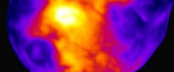
New technique captures activity of an entire brain in a snapshot

When it comes to measuring brain activity, scientists have tools that can take a precise look at a small slice of the brain (less than one cubic millimeter), or a blurred look at a larger area. Now, researchers at Rockefeller University have described a new technique that combines the best of both worlds--it captures a detailed snapshot of global activity in the mouse brain.
"We wanted to develop a technique that would show you the level of activity at the precision of a single neuron, but at the scale of the whole brain," says study author Nicolas Renier, a postdoctoral fellow in the lab of Marc Tessier-Lavigne, professor of the Laboratory of Brain Development and Repair, and president of Rockefeller University.
The new method, described online on May 26 in Cell, takes a picture of all the active neurons in the brain at a specific time. The mouse brain contains dozens of millions of neurons, and a typical image depicts the activity of approximately one million neurons, says Tessier-Lavigne. "The purpose of the technique is to accelerate our understanding of how the brain works."


